Initial Interpretation of Myocardial Perfusion Imaging
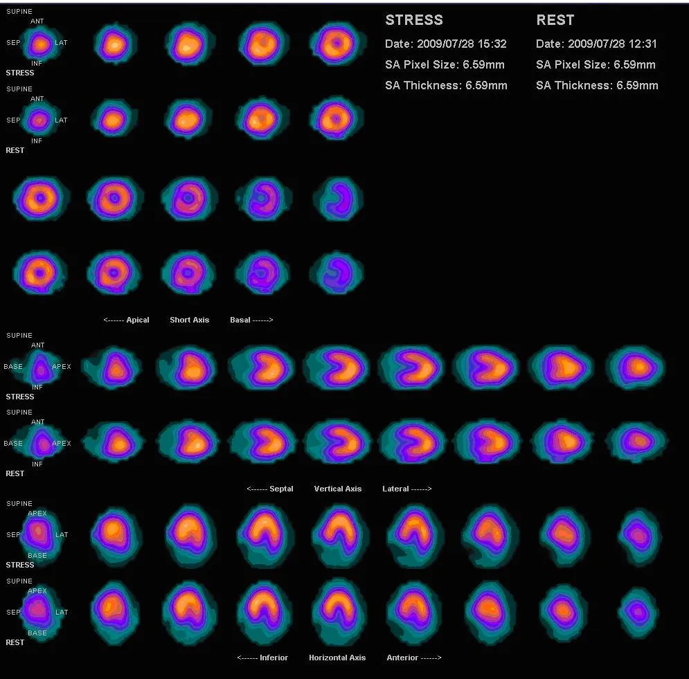
Background knowledge: Coronary steal phenomenon
A stenosed coronary artery receives a smaller proportion of coronary flow than a normal vessel. Distal to the stenosis there will be compensatory vasodilatation to maximize flow to myocardial tissue.
Dipyridamole causes vasodilation of normal coronary arteries, diverting blood flow away from the stenotic vessel. As a result, the cardiac muscle supplied by the stenosed coronary artery becomes ischaemic.
| Myocardial state | Normal | Reversible ischemia | Irreversible ischemia |
| Stress | ↑ perfusion | ↓ perfusion | ↓ perfusion |
| Rest | ↓ perfusion | ↑ perfusion | ↓ perfusion |
Reading
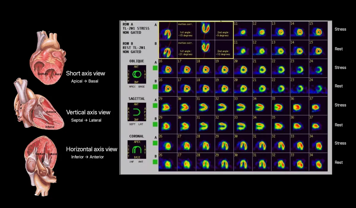
Start with the stress images.
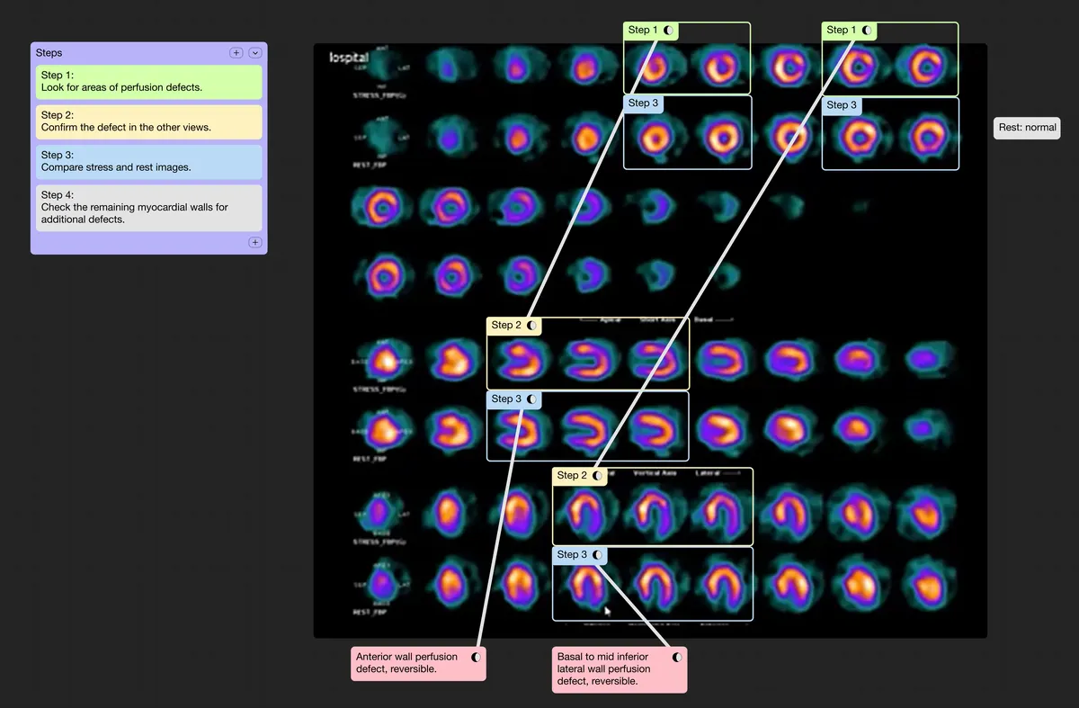
Review the short axis view from apical to mid to basal. Look for areas of perfusion defects.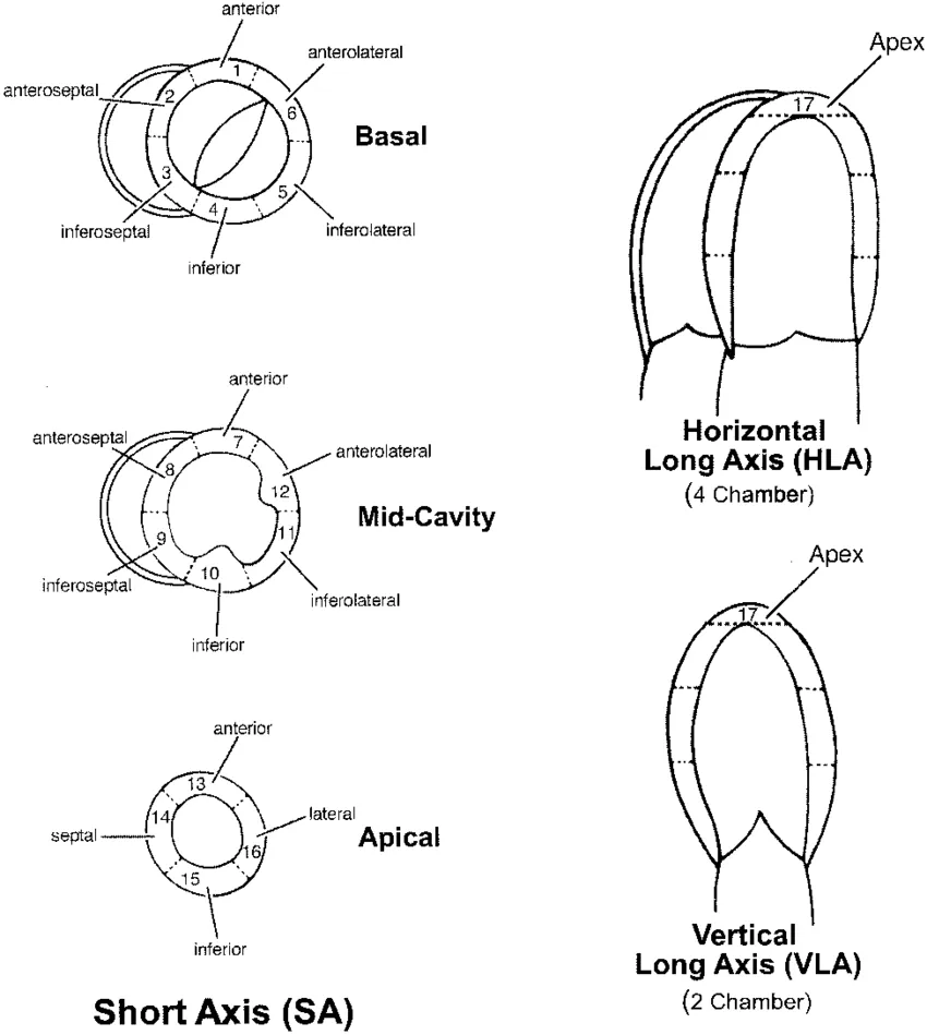
Confirm the defect in the other views.
Correlate the finding with the vertical axis and horizontal long-axis views.Compare stress and rest images.
- Reversible: perfusion defects present on stress but improves at rest.
- Fixed / irreversible: perfusion defects present on both stress and rest.
Check the remaining myocardial walls for additional defects.
Example
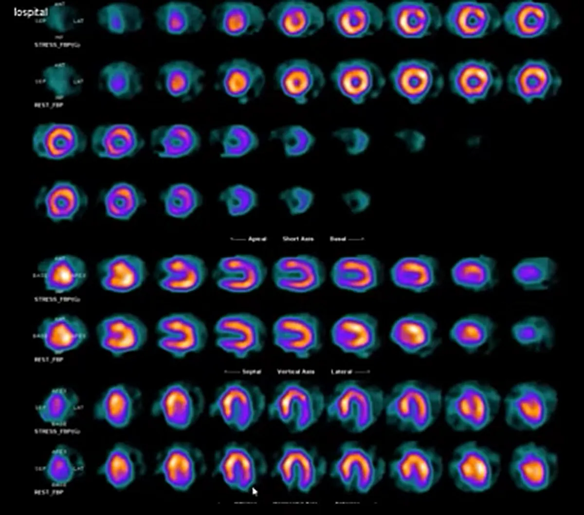
Answer:
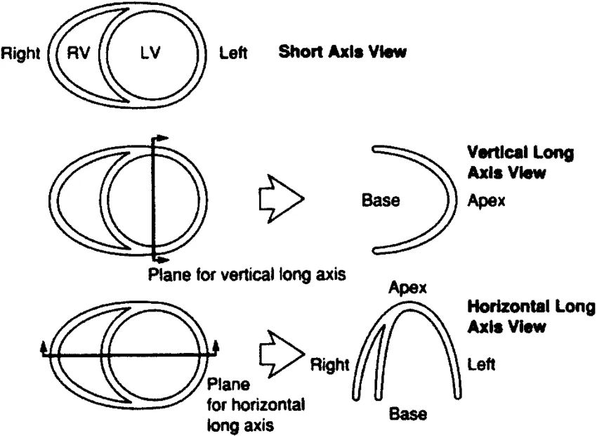
Miscellaneous: How are the axes named?
Reference
Cardiac plane definition and display for tomographic imaging modalities.